
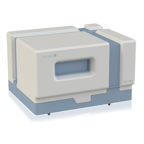
Description

Feature advantages of the CELLAVISTA:

  • Harmonic motion for imaging without agitation of cells during scanning
  • Highly sensitive optics for perfect resolution
  • Up to 5 different magnifications
  • 3 times more sensitive florescence module for shorter exposure times
  • Autofocus performance twice as fast as in previous models
  • Ultrafast multiplex imaging
  • HCS-grade lenses
  • Available in brightfield and with 6 fluorescence channels
  • Faster measurements and less photobleaching than ever before
  • Customisable data and image export function for report creation, documentation and regulatory approval

Applications

These imagers support over 80 imaging algorithms across a wide range of cell based research areas, including:

Cell Line Development

SYNENTEC imaging systems support a range of CLD applications, including Single Cell Cloning, Proof of Monoclonality, CRISPR/Cas9 Tracking, Transfection Efficiency, Confluence Tracking, Cell Viability Monitoring and Fluorescent Activated Single Cell Cloning (FASCC). PAIA applications for protein titre and glycosylation measurements are also supported.

3D Spheroid Imaging

Automated imaging and analysis of 3D spheroids for research and drug discovery applications. Examples include Spheroid Imaging, Spheroid Quantification and Spheroid Live/Dead assays, enabling assessment of spheroid characteristics and viability.

Cell Viability & Toxicity

Assess cell viability and treatment responses using automated brightfield and fluorescence imaging. Applications include AI-based label-free viability, Erythrosin B viability, Trypan Blue viability and LIVE/DEAD toxicity studies. SYNENTEC’s Erythrosin B application provides an alternative to Trypan Blue for high-throughput viable cell counting, with the option to analyse 96 samples in less than five minutes.

Apoptosis & Cell Death

A range of fluorescence-based apoptosis applications can be analysed using automated imaging, including Annexin V, JC-1 and Caspase assays. These assays enable the detection and analysis of different indicators of apoptosis and cell death.

Wound Healing & Cell Migration

Automated time-lapse imaging can be used to monitor wound closure and cell migration throughout an assay. Applications include Wound Healing and Scratch Assays, with image analysis used to quantify changes over time.

iPSC Research

iPSC workflows can be monitored and analysed using applications including iPSC Colony Counting, Fluorescent Pluripotency Studies, Proliferation and Cell Migration, Cell Differentiation Analysis and iPSC-Cell-Marker Characterisation. iPSC colony counting can be used to quantify the number and area of colonies grown on a feeder layer. Together, these capabilities support iPSC-based disease modelling, enabling the study of disease mechanisms, patient-specific phenotypes and therapeutic responses in 2D and 3D cellular models.

Scientific Line

Featuring high-sensitivity 16-bit sCMOS cameras combined with high numerical aperture objectives, the SCIENTIFIC instrument series achieve better resolution in a higher contrast and allow for better detection of weak fluorescent signals. Specifically designed for cancer research and drug discovery applications, the CELLAVISTA SCIENTIFIC achieves remarkable high content imaging in ultra-high throughput.

  • Antibody binding
  • Drug discovery screening
  • Antibody internalisation
  • Image-based predictive modeling
  • High Content Screening

4K Line

The first automated 4K cell imager series with high throughput and configurable resolution settings. Originally launched in 2007, the CELLAVISTA has been continuously developed and is now more powerful than ever. The system is designed for seamless integration into automation platforms and can be easily linked to robotic arms, plate stackers, incubators, liquid handlers and clone pickers (see SYNENTEC’s SYBOT-1000). Coupled with highly sensitive optics for unmatched resolution, the CELLAVISTA 4K is essential for single cell cloning workflows as well as many other high throughput applications.


High content screening using 4K resolution

This image of A549 cells impressively demonstrates the capabilities of SYNENTEC’s CELLAVISTA 4K imager. The actin filaments can be recognized with impressive high definition.

  • Single cell cloning
  • Cell counting and confluency
  • High content assays
  • Localisation studies
  • Translocation studies
  • Predictive modelling and morphological classification

More on SYNENTEC

SYNENTEC’s team of engineers, software developers and biologists have combined their knowledge to develop automated, high throughput cell culture microscopes and fast, easy to use image analysis software. They continuously develop and perfect the imaging systems and work with customers to create bespoke applications to meet their individual cell imaging and analysis needs.

SYNENTEC provide the most versatile cell imagers on the market.

VIEW ALL SYNENTEC PRODUCTS >

Sign up to BPES e-newsletters

Sign Up